Awesome macro photography of what the inside of foam looks like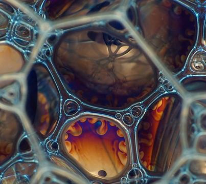
|
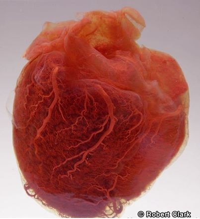
Human heart at the Mütter Museum, Philadelphia Photo by Robert Clark
|
Cardiac muscle with capillaries passing amongst the heart muscle fibers.
Cardiac muscle tissue occurs only in the heart. Its cells are joined end to end. The resulting fibers are branched and interconnected in complex networks. Each cell has a single nucleus. At its end, where it touches another cell, there is a specialized intercellular junction called an intercalated disc, which occurs only in cardiac tissue.
Cardiac muscle is controlled involuntarily and, in fact, can continue to function without being stimulated by nerve impulses. This tissue makes up the bulk of the heart and is responsible for pumping blood through the heart chambers into the blood vessels. 
|
Healthy erythrocyte versus crenated one.
Crenation is caused by dehydration, which distorts the cell. The main function of erythrocytes of course is to distribute oxygen to body tissues and carry the waste carbon dioxide back to the lungs. Crenation leads to severe reduction in their ability to carry out this function.
So drink enough!! Otherwise, your blood will become hyperosmolar and the erythrocytes fluid will be drained out, which makes them look like this. Same happens if you drink salt water. Don't drink salt water! (such valuable tipps, ain't they? Magnification x6200, by Steve Gschmeissner 
|
Incredible photograph of the arteries and veins of the human headPhoto by James Bareham, photographer of the New York based creative studio The New Cruelty

|
A human fetus in the mother's wombThis image was created for a National Geographic special called In The Womb by Producer Peter Chinn.
He used a combination of ultrasound technology, tiny cameras, and computer design to create these incredible images that replicate what a fetal human being looks like. This is an incredible window into the womb, laying bare the mysteries of the beginning of life! 
|
Scanning electron micrograph of the inside of a cancer cell.
This cell originates from a squamous cell carcinoma, a type of skin cancer. The cell has been frozen and split open to reveal its nucleus.
Author: Anne Weston http://www.cellimagelibrary.org/ 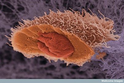
|
Marvelous photograph of a majestic elephant in Botswana by Ben Cranke.
Male african elephants are the largest surviving terrestrial mammals. Unfortunately they get killed every day for their ivory and there were recent prognoses that in ten years there may be no wild living elephants anymore.

|
Awesome micrograph of a nerveIn this transmission micrograph you can see a myelinated nerve fibre (axon) from the lining of the trachea (windpipe). Myelin is an insulating fatty layer that surrounds the nerve fibre, increasing the speed at which nerve impulses travel.
It is formed when Schwann cells wrap around the fibre, depositing layers of myelin between each coil. The outermost layer consists of the Schwann cell's cytoplasm and is known as the neurolemma or sheath of Schwann. Magnification x4000, by Science photo library. 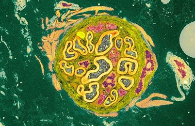
|
This is what a tree looks like when it's dancing

|
This macro shot of a ladybird covered in beads of morning dew was captured by German photographer Jens Kolk in his back garden in Potsdam.

|
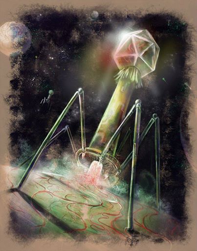
Beautiful bacteriophage illustration
|